Researchers have developed a simplified process that could enhance personalization of cancer therapy based on a single nuclear medicine scan. The novel method uses a follow-up single photon emission computed tomography/computed tomography (SPECT/CT) scan to obtain reliable radiation dose estimates to tumors and at-risk organs. The study is published in The Journal of Nuclear Medicine.
“In most radionuclide therapies, a fixed amount of radionuclide is administered, which does not take into account individual patient variability in tumor and normal tissue uptake, and hence absorbed dose,” said John Violet, BSc, MBBS, MRCP, FRCR, Ph.D., FRANZCR, radiation oncologist at the Peter MacCallum Cancer Centre in Melbourne, Australia. “‘Dosimetry-led’ administration, on the other hand, where administered activity is adjusted based upon predictable tumor and normal tissue absorbed doses, has the potential to greatly improve therapeutic outcomes. As dosimetry requires multiple SPECT/CT scans post-therapy, however, it is rarely performed outside clinical trials.”
Rather than rely on multiple SPECT/CT scans to calculate dosimetry, researchers sought to develop a dose-response estimation from a single post-treatment scan. The study presented a simplified dosimetry method and applied it to the assessment of 177Lu-PSMA-617 therapy for metastatic prostate cancer. 177Lu-PSMA-617 therapy is known to be a highly active therapy for PSMA-expressing metastatic prostate cancers, but currently there is no standardization of how treatment is given.
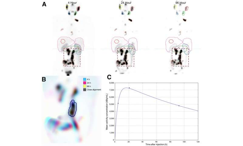
In the study, 29 patients receiving 177Lu-PSMA-617 were imaged with SPECT/CT at four, 24 and 96 hours to characterize tracer pharmacokinetics in tumors and in normal tissues of interest. To evaluate serial measurements, three-dimensional volumes of interest were defined on the first image volume and a method was developed to automatically align each contoured subregion and assess pharmacokinetics. This data was used to develop a population PSMA clearance model for tumor and healthy organs.
By applying a generic pharmacokinetic model for PSMA retention, it is possible to estimate radiation absorbed dose with a single image measurement. The range of added uncertainty can be appreciated based on the variability in population curves when normalized to the single time point. Tumor dose estimates were most accurate using delayed scanning at times beyond 72 hours. Dose to healthy tissues was best characterized by scanning patients in the first two days of treatment because of the larger degree of tracer clearance in this early phase.
“This work shows for the first time that following Lu-PSMA administration, it is possible to determine a good estimate of activity and absorbed dose with just a single measurement,” noted Violet. “Simplification of dosimetry methods to make them a routine application would greatly assist in developing radiobiological models specific to Lu-PSMA, and other radionuclide therapies, allowing optimization of therapy for individual patients that are maximally effective and safe.”
Source: Read Full Article