In a recent study published in the Emerging Infectious Diseases Journal, researchers analyzed epidemiologic and phylogeographic characteristics of the highly pathogenic avian influenza virus (HPAI H5N1) viral organisms detected globally between 2020 and 2022.
The study also investigated the pathogenicity of H5N1 viruses among waterfowl hosts and mammals. They also evaluated the effectiveness of the currently used H5/H7 trivalent inactivated vaccine against H5N1 viruses.
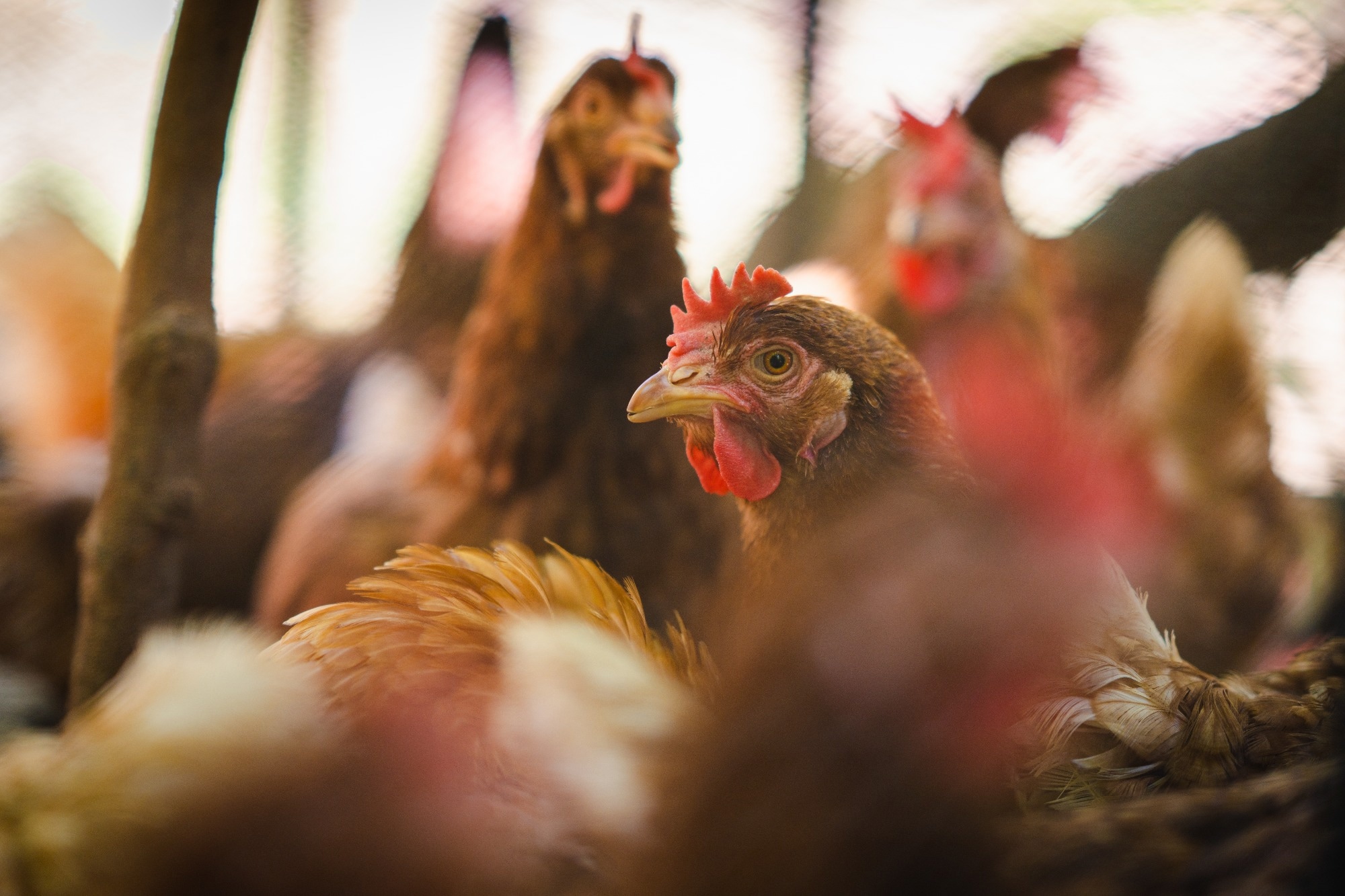
 Study: Highly Pathogenic Avian Influenza Virus (H5N1) Clade 2.3.4.4b Introduced by Wild Birds, China, 2021. Image Credit: WassanaPanapute/Shutterstock.com
Background
Clade 2.3.4.4b H5N8 viral transmission led to widespread outbreaks in wild birds and poultry in Asia, Africa, and Europe, between January 2020 and October 2021. During the period, the virus underwent reassortments in different regions and nations, generating H5N1, H5N2, H5N3, H5N4, H5N5, and H5N6 viruses that comprise the hemagglutinin (HA) gene of the 2.3.4.4b clade.
Among novel subtypes, only the H5N1 virus has been extensively transmitted through migratory avian species, resulting in outbreaks in Africa, Europe, North America, and Asia since its emergence in the Netherlands in October 2020.
The viruses have also infected humans and several marine and carnivorous mammals, raising health concerns for poultry and humans globally.
About the study
In the present study, researchers evaluated the epidemiology, genetic evolution, transmission patterns, and pathogenicity of clade 2.3.4.4b H5N1 viruses and the H5-Re14 vaccine’s immunogenicity against H5N1 viruses.
Between 2020 and 2021, the team obtained 507 swab specimens, 7,421 fecal specimens, and six tissue specimen samples from undomesticated avian species and amplified them in chick embryos.
Subsequently, hemagglutinin inhibition (HI) tests were performed using H1 to H16 reference sera to identify their HA subtypes, and reverse transcription polymerase chain reaction (RT-PCR) was performed to verify neuraminidase (NA) gene subtypes using N1 to N9 primers.
Viral ribonucleic acid (RNA) was extracted from the H5N1-positive chicken embryos, and the hosts were identified using deoxyribonucleic acid (DNA) barcodes and the cytochrome C (Cyt C) oxidase I gene.
The team downloaded genetic data obtained between 1 January 2020 and 17 October 2022 from the Global Initiative on Sharing All Influenza Data (GISAD) database and aligned genetic sequences to determine sequence identity and construct the maximum likelihood tree.
Further, the patterns of transmission for 2.3.4.4b clade H5N1 viral transmission were investigated. Sequences of 1,529 viral isolates were grouped into 11 different geographical groups, and the diffusion rates were ascertained using Bayesian search selection.
Further, A/whooper swan/Henan/14/2021 (WS/HeN/14/2021) and A/mandarin duck/Heilongjiang/HL-1/2021 (MD/HLJ/HL-1/2021) isolates were tested in ducks and mice. BALB/c mice were inoculated intranasally with H5N1’s 101 to 106 50% egg infective dose (EID50) and monitored over two weeks to calculate the 50% lethal dose (LD50).
The mice were euthanized three days post-inoculation to determine H5N1 titers in the murine brains, lungs, nasal turbinates, spleens, and kidneys. Likewise, eight ducks were intranasally inoculated with 106 EID50 of the virus, and their body tissues were examined to assess H5N1 replication.
In addition, cloacal and oropharyngeal swabs were obtained two weeks post-inoculation to evaluate H5N1 shedding. For the antigenic analysis, HI was performed, and chicken antisera of H5-Re11, 12, 13, and 14 viruses were generated.
For the 2.3.4.4b clade H5N1 virus challenge study, chickens were vaccinated intramuscularly with the trivalent vaccine and inoculated with MD/HLJ/HL-1/2021’s 105 egg 50% infective dose. Their cloacal and oropharyngeal swabs were obtained to detect H5N1.
Results
As a component of daily Centres for Disease Prevention and Control (CDC) surveillance of undomesticated avian species between 2020 and 2021, 7,934 specimens were obtained, among which 17 HPAI H5N1 viral organisms and 20 low pathogenic avian influenza viruses (LPAI) of several subtypes were identified. HPAI viruses’ HA and NA genes comprised 98% to 100% similar nucleotides.
The phylogenetic tree for the 2.3.4.4b clade H5 strains’ HA genes branched into the Africa/Eurasia group (n=15) and the East Asia group (n=2). The neuraminidase genes of all 17 HPAI viruses were grouped with H5N1 viruses detected in Europe in 2020.
Nucleotide-level sequence identity ranged between 93.0% and 100.0% for H5N1 viruses’ matrix (M), polymerase acidic (PA), polymerase basic (PB) 1, PB2, non-structural protein (NS), and nucleoprotein (NP) genes. The 2.3.4.4b clade H5N1 viruses’ genomic diversity extended beyond the NA and HA genes.
Notably, eight genes from the mandarin duck isolates of 2020 and 2021 and the remaining 15 HPAI viral organisms were identical. Two genotypes were identified for the HPAI viruses: two H5N1 viruses detected in Heilongjiang were of the G07 genotype, whereas the remaining 15 were of the G10 genotype.
The findings indicated that the 2.3.4.4b clade H5N1 viral organisms had undergone complicated reassortments with LPAI viral organisms in several locations from 2020 onward. The viruses of both genotypes propagated among domestic ducks in China two months after being identified among wild birds.
The researchers discovered 22 virus transmission pathways, and Europe was the epicenter of the worldwide 2.3.4.4b clade H5N1 viral proliferation. The HA genes of the viruses comprised the 137A and 192I residues that increased their affinity for human receptors.
The team detected whooper swan and mandarin duck strains in murine lungs, nasal turbinates, kidneys, and spleens. The viruses showed moderate pathogenicity in mice but were lethal to ducks. The H5/H7 trivalent vaccine protected the animals robustly against the H5N1 viruses.
Conclusion
Overall, the study findings highlighted the evolutionary, transmission, and infection dynamics of 2.3.4.4b clade H5N1 viral organisms, underscoring the need for increased H5N1 surveillance and underpinning H5/H7 trivalent vaccinations to protect poultry from H5N1 infections.
-
Tian J, et al. (2023) Highly pathogenic avian influenza virus (H5N1) clade 2.3.4.4b introduced by wild birds, China 2021. Emerg Infect Dis., doi: 10.3201/eid2907.221149. https://wwwnc.cdc.gov/eid/article/29/7/22-1149_article
Posted in: Medical Science News | Medical Research News | Disease/Infection News
Tags: Avian Influenza, DNA, Epidemiology, Evolution, Gene, Genes, Genetic, Genomic, H5N1, Infectious Diseases, Influenza, Lungs, Nucleotide, Nucleotides, Polymerase, Polymerase Chain Reaction, Proliferation, Protein, Ribonucleic Acid, RNA, Structural Protein, Transcription, Vaccine, Virus

Written by
Pooja Toshniwal Paharia
Dr. based clinical-radiological diagnosis and management of oral lesions and conditions and associated maxillofacial disorders.
Source: Read Full Article