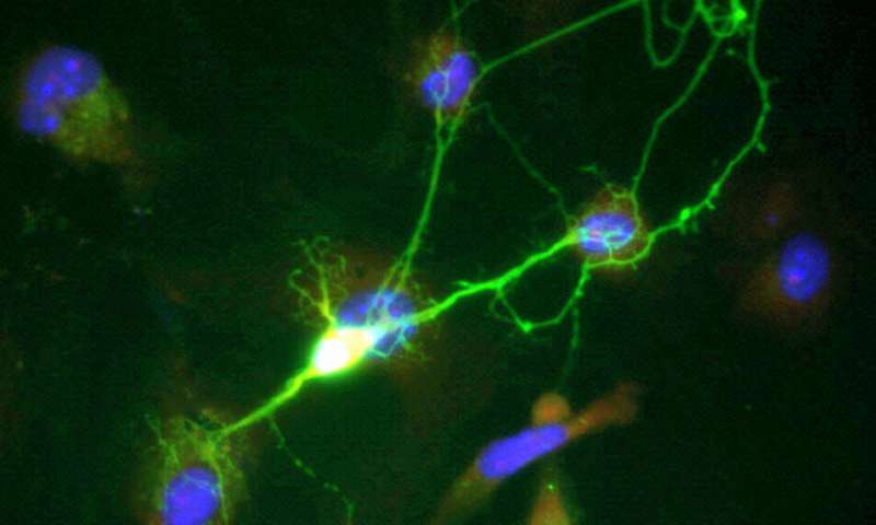
Depression, a common mental disorder, can severely disturb the life of its patients. A hypothesis stated that the excitatory synapses characterized by the post-synaptic density (PSD) in the hypothalamus, which played a crucial role in emotion regulation, may be involved in the pathogenesis of depression. However, how synaptic regulation in the hypothalamus has influence on depression has not been fully understood.
In a study published in Acta Neuropathologica, a group led by Prof. Zhou Jiangning from the University of Science and Technology of China of the Chinese Academy of Sciences revealed the excitatory synaptic regulation of corticotropin-releasing hormone (CRH) neuron in the hypothalamus, and that the excitatory synaptic regulation plays an important role in the depression pathology. The researchers showed the mechanism of involvement of synaptic-associated protein PSD-93 in the pathogenesis of depression.
CRH neuron in the hypothalamic paraventricular nucleus (PVN) is the driving force in hypothalamic-pituitary-adrenal (HPA) axis regulation. The researchers observed that an increased density of PDS-93-CRH co-localized neurons in the PVN of patients with major depression disorder (MDD). Subsequently, PSD-93 in CRH neurons was locally overexpressed by virus injection.
The results showed that overexpression of PSD-93 led to depression-like behaviors in mice and increased excitatory synaptic activity of CRH neurons, while PSD-93 knockdown could decrease excitatory synaptic activity, and alleviate depression-like behaviors in a lipopolysaccharide (LPS)-induced depression model.
Using fiber photometry recordings in vivo, the researchers found that the release of glutamate from activated microglia to CRH neurons was significantly increased after LPS treatment, which induced depression-like behavior. The results of fluorescence in situ hybridization (FISH) experiments indicated that LPS activated microglia and increased the synthesis of glutaminase in microglia.
Finally, the researchers studied postmortem brain samples of patients with MDD through immunohistochemistry and immunofluorescence staining, and they observed an increase in the density of PSD-93 and CRH co-expression neurons in the hypothalamic PVN.
Source: Read Full Article