An interesting new paper presents a new assay for B and T lymphocytes in individuals infected with the severe acute respiratory syndrome coronavirus 2 (SARS-CoV-2). This B And T cell Tandem Lymphocyte Evaluation (BATTLE) assay allows for the simultaneous identification of reactive T and B cells, thus sparing sample use for other important purposes.
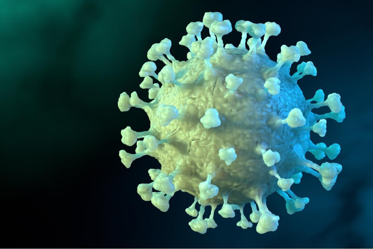
 Study: Simultaneous analysis of antigen-specific B and T cells after SARS-CoV-2 infection and vaccination. Image Credit: CROCOTHERY/Shutterstock
A preprint version of the study is available on the bioRxiv* server while the article undergoes peer review.
Background
The immune background of this infection has been under severe scrutiny to unravel its nature, intensity, and characteristics. This involves the study of T and B lymphocytes that play distinct but irreplaceable roles in the evolution of immunity against the virus or its vaccines.
Currently, identification of T and B cells that are specifically reactive to the virus involves measuring the number of activated functional cytokine-producing T cells, finding evidence of intracellular cytokine generation on stimulation with appropriate peptides, the use of peptide/HLA probes, and the detection of activation markers after antigen recognition and binding.
Both these approaches involve using separate samples for the study of T and B cells. The need to conserve resources by examining both types of cells in parallel led to the current experimental verification of this assay. The purpose was to identify and phenotype B cells, CD4+ and CD8+ T cells, thus saving the sample and allowing the responses of these two types of cells to the same antigen to be tracked dynamically over time.
What did the study show?
The assay was tested in a large cohort of recovered individuals who had donated convalescent plasma after symptomatic mild COVID-19, AT 30-75 days. This period is expected to correspond to antigen-specific reactive memory lymphocytes before natural infection but after the inflammatory response has resolved, thus excluding evidence of acute responses and preserving long-term memory responses.
All participants had obtained two doses of an mRNA vaccine, with at least nine months between the last negative test for the virus and the next blood test, but within 22 weeks following vaccination.
The researchers used an optimized Activation-Induced Marker (AIM) assay to pick up reactive T cells stimulated by pools of SARS-CoV-2 spike peptides, overlapping with each other, and spike reactive B cells with pike trimer fluorescent tetramers. They tracked B and T cells populations over time in individuals who had been first infected naturally by the virus and then vaccinated.
The BATTLE assay proved to be efficient at quantifying and isolating both B and T cells reactive to the spike antigen simultaneously, using one sample of peripheral blood cells. The results show that mRNA vaccines stimulate both B and CD8+T cells. However, CD4+ T cells respond to vaccine stimulation only when the T cells were undetectable or very low after natural infection.
Independent serologic profiling showed that anti-nucleocapsid (anti-N) immunoglobulin G (IgG) titers declined between first and second vaccine doses. Anti-N IgG is a sign of natural infection, but not of vaccination since the N antigen is not included in this type of vaccine. Anti-N IgM was mostly undetectable.
The anti-spike receptor-binding domain (RBD) IgG responses also increased with vaccination, but not IgM. IgG, but not IgM, antibodies to the Alpha and Beta variant spike RBDs were also found to rise after mRNA vaccination, to different degrees.
Vaccination led to a rise in spike-reactive B cells elicited by natural infection, both in frequency and number. However, these were not found in samples collected before the pandemic began. CD8+ and CD4+ cells showed divergent responses with the T cells. The latter showed a slight increase in CD8+ T cells reactive to the spike after vaccination, but not for non-activated CD8+ T cells.
Thus, B and CD8+ T cells were found to show a coordinated response against the RBD, but not the CD4+ T cells. Spike-reactive B cell responses were correlated with serum IgG titers to the RBD for all tested variants.
These data suggest that mRNA vaccination of previously-infected individuals influences T cell subsets asymmetrically, and may allow those who mount a sub-optimal response to infection to achieve an immunological setpoint not reached via natural infection.”
What are the implications?
B cells and both subsets of T cells can be identified and quantified with the same sample. This shows that perhaps spike-reactive B and T cells are enhanced by mRNA vaccines following natural infection, but not to an equal extent. Adding more antigens to the vaccine could improve the memory immune response.
Yet, despite the divergent trends, the contraction and expansion of specific subsets can be seen with these results, with B cells expanding more in response to the vaccine than spike-reactive CD4+ T cells. Perhaps, B and T cells have distinct immunological setpoints after immune stimulation. This could lead to a compartment-specific immune memory response which enhances protective immunity.
Our findings suggest divergent patterns of B and T cell subset mobilization with repeated antigen encounter and highlight the utility of examining these populations in concert.”
The findings could help expand the array of diagnostic tests using B and T cells. The researchers plan to use BATTLE to explore the adaptive immune responses to more antigens in the future.
*Important notice
bioRxiv publishes preliminary scientific reports that are not peer-reviewed and, therefore, should not be regarded as conclusive, guide clinical practice/health-related behavior, or treated as established information.
-
Newell, K. et al. (2021) "Simultaneous analysis of antigen-specific B and T cells after SARS-CoV-2 infection and vaccination". bioRxiv. doi: 10.1101/2021.12.08.471684. https://www.biorxiv.org/content/10.1101/2021.12.08.471684v1
Posted in: Medical Science News | Medical Research News | Disease/Infection News
Tags: Antibodies, Antigen, Assay, B Cell, Blood, Blood Test, CD4, Cell, Convalescent Plasma, Coronavirus, Coronavirus Disease COVID-19, Cytokine, Diagnostic, Evolution, Frequency, Immune Response, immunity, Immunoglobulin, Intracellular, Lymphocyte, Pandemic, Peptides, Phenotype, Receptor, Respiratory, SARS, SARS-CoV-2, Severe Acute Respiratory, Severe Acute Respiratory Syndrome, Syndrome, Vaccine, Virus

Written by
Dr. Liji Thomas
Dr. Liji Thomas is an OB-GYN, who graduated from the Government Medical College, University of Calicut, Kerala, in 2001. Liji practiced as a full-time consultant in obstetrics/gynecology in a private hospital for a few years following her graduation. She has counseled hundreds of patients facing issues from pregnancy-related problems and infertility, and has been in charge of over 2,000 deliveries, striving always to achieve a normal delivery rather than operative.
Source: Read Full Article