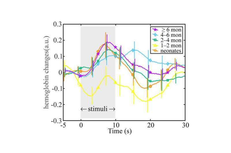
Researchers from Tokyo Metropolitan University have measured how oxygenated hemoglobin levels in the blood change in infants’ brains in response to touch. Using spectroscopy methods with external sensors placed on the scalp of sleeping infants, they found that the time at which levels peak doesn’t change with infant age, but the amount by which it varies over time does. Insights like this shed light on how the physiology of infants develops.
The first phase of a newborn’s life is a dazzling array of rapid developmental changes. And neuroscience is giving us unprecedented insight into these processes, powered by state-of-the-art measurement techniques. Two key tools on this journey are functional magnetic resonance imaging (fMRI) and near-infrared spectroscopy (NIRS) of the brain.
With NIRS, an array of external sensors placed on an infant’s head lets scientists track how the flow of different compounds in the brain changes over time. There is a particular focus on hemoglobin, the key oxygen-carrying component of our blood. As the brain responds to input from the outside world like light, heat, and touch, oxygen rushes to the brain; NIRS lets us pick out levels of hemoglobin, even telling apart whether it is carrying oxygen or has already delivered its cargo.
Now, a team led by Assistant Professor Yutaka Fuchino from Tokyo Metropolitan University has applied NIRS to study how infant brains respond to touch. The study is published in the journal NeuroImage.
Looking at a range of infants aged zero to one, the team placed sensors on infants’ scalps as they slept, and tracked how oxygenated hemoglobin levels changed over time as their limbs were given a very gentle shake. Interestingly, infants at different ages over their first year showed a very similar response time, with a small peak in hemoglobin appearing a few minutes after the stimulus began.
The fact that infants in their first month responded in basically the same way as those over six months suggests that the factors determining the speed of the response are complete at birth.
However, they also noticed that the range over which the signal varied, or the amplitude, was markedly different between infants in different age groups. The change was also not linear, in that it dipped for infants one to two months old then increased again as the age increased.
This curious behavior could be attributed to certain aspects of a newborn infant’s first few months. For one, when they are born, there is a rapid rise in oxygen levels due to being able to breathe normally for the first time. This inhibits the production of erythropoietin, a small protein responsible for stimulating red blood cell production; this leads to temporary anemia over the first few months.
Levels gradually recover as erythropoietin production ramps up again to compensate. Other factors include developments in nerves, veins, and levels of blood flow.
Importantly, the team’s work highlights how NIRS statistics of hemoglobin levels in the brain can reflect developmental changes. Future studies promise to compare other physiological responses to changes in blood flow dynamics, yielding further insights into how the brain and its interactions with the body develop.
More information:
Yutaka Fuchino et al, Developmental changes in neonatal hemodynamics during tactile stimulation using whole-head functional near-infrared spectroscopy, NeuroImage (2023). DOI: 10.1016/j.neuroimage.2023.120465
Journal information:
NeuroImage
Source: Read Full Article